


In order to receive better information about their external environment, blind individuals compensate their lack of visual input by responding to somatosensory or auditory input with improved sensitivity and accuracy. The brain can adapt to sensory deprivation in one modality (e.g. visual cortex) by increasing plasticity and retuning neuronal circuits in other remaining modalities (cross-modal plasticity). Although dynamic cortical reorganisation among different sensory areas is critical for this adaptation, the molecular and cellular mechanisms underlying it are still poorly understood. In the present study, Professor Takuya Takahashi and his colleagues at Yokohama City University (and Albert Einstein College of Medicine, NY, USA) present a comprehensive study that explains how visual loss leads to improved somatosensory function in the rat on a molecular, cellular, and circuit level. Understanding the molecular mechanism through which cross-modal plasticity is established will further our knowledge of how to selectively restore or retune neuronal circuits in individuals that suffered from sensory dysfunction.
This study is published in the February issue of Neuron.
This study was mainly funded by "Development of biomarker candidates for social behaviour" carried out under the Strategic Research Program for Brain Sciences by the Ministry of Education, Culture, Sports, Science and Technology of Japan, and Special Coordination Funds for Promoting Science and Technology.
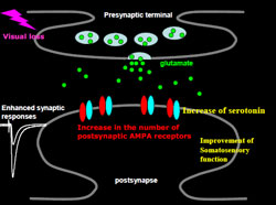
The majority of excitatory synapses in the mammalian central nervous system use glutamate as a neurotransmitter. Fast glutamatergic transmission is mainly mediated by AMPA (ƒ¿-amino-3-hydroxy- 5-methyl-4-isoxazole propionic acid) type ionotropic glutamate receptors (AMPARs) (Figure). AMPARs are tetramers comprised of a combinatorial assembly of four subunits, GluR1-4. Synaptic plasticity at glutamatergic synapses is considered to be crucial for cognitive functions such as learning and memory. The best-studied form of synaptic plasticity is LTP (Long-term potentiation), and its molecular mechanisms have been extensively characterised. Synaptic delivery and addition of AMPARs appears to be a major mechanism regulating postsynaptic expression of LTP. LTP inducing stimuli in vitro drive GluR1-containing AMPARs into synapses and enhance synaptic transmission. The same molecular modification occurs during experience-dependent neuronal reorganisation in vivo (Figure). Sensory experience early in development delivers GluR1 into synapses of the rodent barrel cortex which receives input from whiskers. Fear learning also induces synaptic GluR1 delivery in amygdala.
Although blind human often possess other senses with extraordinary ability, the molecular and cellular mechanisms underlying it is still poorly understood. In this study, Professor Takuya Takahashi and his colleagues present a comprehensive study that explains how visual loss leads to improved whisker function in the rat on a molecular, cellular, and circuit level. They found that visual loss increased the extracellular serotonin levels in the barrel cortex, thereby facilitating the delivery of GluR1 into layer 4-2/3 synapses. Furthermore, this resulted in a sharpening of the functional whisker-barrel map. Thus, serotonin mediates cross-modal cortical reorganisation induced by sensory deprivation through the facilitation of synaptic GluR1 delivery and results in the improvement of remaining sensory functions (see illustration).
This study presents the molecular and cellular mechanisms underlying functional compensation of sensory loss. These findings could lead to the invention of novel therapy to improve the performance during rehabilitation of patients with sensory loss.
Source: Yokohama City University


